Table of Contents
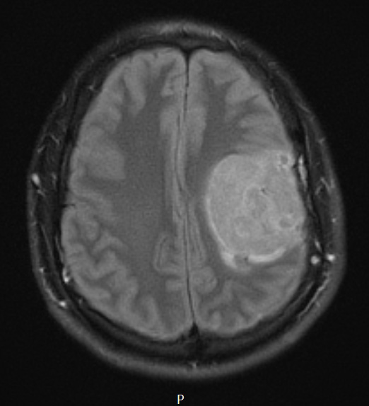
Washington University Experience | NEOPLASMS (GLIAL) | Pleomorphic Xanthoastrocytoma (PXA) | 17A1 PXA Anaplastic WHO III (Case 17) TIRM - Copy
Case 17 History ---- The patient is a 22-year-old man who initially presented in January 2014 with seizures and right facial weakness. MR imaging performed at that time showed a 2.4 x 2 cm, T1 hyperintense, T2 heterogeneous, posterior left frontal lobe hemorrhagic mass with associated mass effect. Over the latter part of January into April 2014, the lesion decreased in size in conjunction with evolving hematoma. MR imaging performed in December 2014 showed an interval marked increase in the left frontal lobe lesion up to 5.2 cm, which showed both cystic and lobulated enhancing solid component and approximately 9 mm of left to right midline shift. Operative procedure: Left frontal craniotomy for resection of tumor with use of intraoperative stereotactic neuronavigation. --- The lesion is hyperintense on TIRM scan.

