Table of Contents
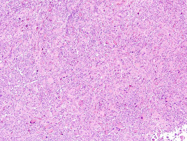
Washington University Experience | NEOPLASMS (GLIAL) | Pleomorphic Xanthoastrocytoma (PXA) | 17B2 PXA Anaplastic WHO III (Case 17) H&E 6.jpg
17B2-6 Sections show a tumor consisting of epithelioid-rhabdoid-gemistocytic-vacuolated-xanthomatous cells growing in a vaguely fascicular pattern. Some areas appear well-demarcated from the adjacent parenchyma, while others show clear infiltration. Macrocysts are present. There is significant nuclear pleomorphism consisting of round-to-ovoid morphology, moderately irregular contours, marked variation in nuclear size, coarsely clumped chromatin, prominent nucleoli, intranuclear inclusions, and frequent multinucleation. Eosinophilic granular bodies are readily identified and in some areas are numerous. Finally, there is focal parenchymal and perivascular chronic inflammation.

