Table of Contents
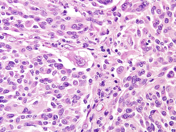
Washington University Experience | NEOPLASMS (GLIAL) | Pleomorphic Xanthoastrocytoma (PXA) | 18A8 Pleomorphic Xanthoastrocytoma (Case 18) H&E 22
Some of the tumor cells are multinucleated and bizarre. However, mitotic figures are hard to find and there is no evidence of necrosis or endothelial proliferation. ---- Ancillary data (not shown): Reticulin highlights increased deposition around groups of cells focally. A subset of tumor cells display strong immunoreactivity for GFAP. The majority of tumor cells are negative for synaptophysin, although there is a high degree of background staining. The Ki-67 labeling index appears low in most areas of the tumor. Focally, it is increased to roughly 6.7%; however, many of the immunoreactive cells are small and rounded, potentially representing lymphocytes. Only rare nuclear positivity is seen with a p53 protein stain. ---- Comment: The morphologic, histochemical, and immunohistochemical features are consistent with the diagnosis of pleomorphic xanthoastrocytoma, WHO grade 2.

