Table of Contents
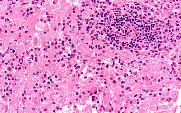
Washington University Experience | NEOPLASMS (GLIAL) | Pleomorphic Xanthoastrocytoma (PXA) | 19A1 (Case 19) H&E 40X 2
Case 19 History ---- The patient is a 19 year old man with a history of NF1. Imaging studies recently demonstrated an enhancing right parieto-occipital mass that grew over subsequent imaging. Therefore, a resection was performed. ---- 19A1-8 Sections reveal a solid and focally infiltrative appearing neoplasm with perivascular lymphocytic cuffing. There is moderate to marked nuclear pleomorphism, with most of the tumor cells having an astrocytic appearance. They contain oval to irregular hyperchromatic nuclei with abundant eosinophilic cytoplasm arranged in either an epithelioid or spindled fashion.

