Table of Contents
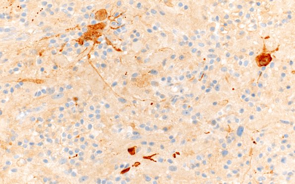
Washington University Experience | NEOPLASMS (GLIAL) | Pleomorphic Xanthoastrocytoma (PXA) | 19C2 (Case 19) SYN 40X
A few tumor cells show cytoplasmic immunoreactivity for synaptophysin. (SYN IHC) ---- Ancillary studies (not shown): A reticulin stain shows increased deposition around blood vessels and focally increased deposition between tumor cells. The neurofilament protein stain shows a solid and focally infiltrative growth pattern, the latter containing entrapped
immunoreactive axons. There is strong positivity for neuron specific enolase. There are scattered CD45 positive lymphocytes and CD138 plasma cells. The CD34 stain highlights blood vessels and rare tumor cells. The Ki-67 labeling index is low in the tumor cells. A histochemical stain for PAS with diastase reveals rare eosinophilic granular bodies. ---- Comment: The morphologic, histochemical and immunohistochemical features are consistent with the diagnosis of pleomorphic xanthoastrocytoma, WHO grade 2. It is well recognized that rare neuronal elements may also be seen in pleomorphic xanthoastrocytoma and the presence of xanthomatous astrocytes in this tumor favor this interpretation, rather than ganglioglioma. The finding of a pleomorphic xanthoastrocytoma in the setting of NF1 is unusual, but has been reported.

