Table of Contents
Washington University Experience | NEOPLASMS (GLIAL) | Pleomorphic Xanthoastrocytoma (PXA) | 1B2 Pleomorphic Xanthoastrocytoma (Case 1) H&E 2
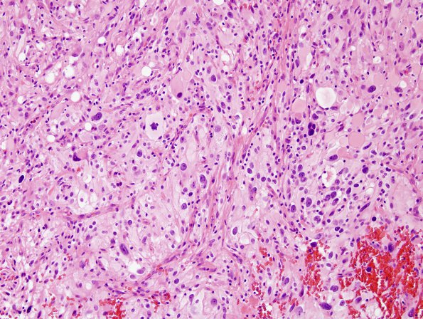
1B2-8 Sections show a cellular glial cell neoplasm composed of sheets of pleomorphic cells. While the majority of cells have a spindled cytoplasm with elongated nuclei, some cells have enlarged nuclei and an irregular nuclear border. Other cells have a more epithelioid appearance and in some of these a xanthomatous appearance is seen. Occasional mitoses are identified. There is no evidence of necrosis. Small numbers of eosinophilic granular bodies (EGBs) are also seen scattered throughout the tissue.

