Table of Contents
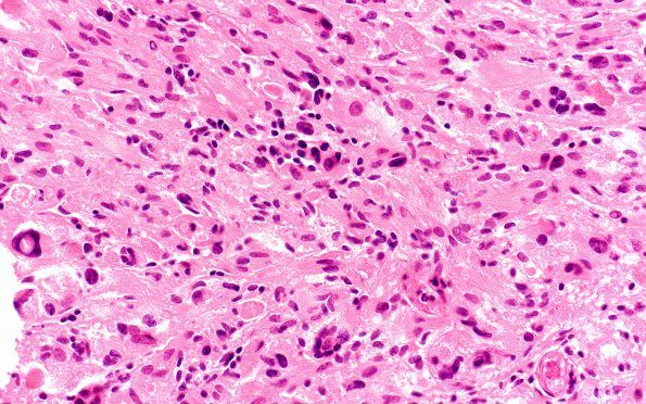
Washington University Experience | NEOPLASMS (GLIAL) | Pleomorphic Xanthoastrocytoma (PXA) | 21A1 (Case 21) H&E 40X 2
Case 21 History ---- The patient is a 28 year old man with 20 year history of complete partial seizures. MRI showed an enhancing lesion in the right parahippocampal and fusiform gyri. Clinical diagnosis: Brain tumor. Operative procedure: Craniotomy, right, for tumor excision with intraoperative mapping.
21A1-3 Examination of the sections show a pleomorphic xanthoastrocytoma. The tumor is composed of markedly pleomorphic nucleated cells with abundant cytoplasm arranged in sheets and strands as well as spindled astrocytes. The tumor cells show marked pleomorphic hyperchromatic nuclei, some with prominent nucleoli or multinucleation. The tumor displays a marked vascularity. There is no evidence of mitotic activity, endothelial hyperplasia, or necrosis within the tumor. There is a perivascular lymphocellular infiltrate with scattered lymphoplasmacytic cells between the tumor cells.

