Table of Contents
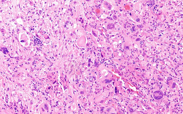
Washington University Experience | NEOPLASMS (GLIAL) | Pleomorphic Xanthoastrocytoma (PXA) | 3A1 PXA, anaplastic (Case 3) H&E 7
Case 3 History ---- The patient is a 58-year-old man with history of atypical central neurocytoma of the left lateral ventricle, status post resection in 2003 , and, more recently, melanoma of the left leg, status post resection at an outside institution in September 2017. Recently, he presented with a history of reduced visual acuity over 2-3 years, dizziness for 3 months, headache for 2 months, and ataxia for 1 month. Head CT at an outside institution showed a 5.7 cm left temporal lobe mass suspicious for malignancy. He received two doses of dexamethasone without improvement in his symptoms. Brain MRI shows a 5 x 4.9 x 3.4 cm solitary rim enhancing lesion in the left temporal lobe with mixed solid and cystic components, and a rim of slight diffusion restriction, without internal diffusion restriction. Radiological impression: high grade glial neoplasm; metastases is less likely given a solitary lesion. Operative procedure: Left craniotomy for tumor excision. ---- 3A1-4- Many cells are very large, with abundant variably pale eosinophilic cytoplasm, multiple highly atypical, often bizarre, nuclei and prominent nucleoli; others are small and variably polygonal or spindled, with modest eosinophilic cytoplasm, and atypical nuclei. They also appear in pavemented sheets and fascicles. Accompanying these tumor cells are occasional clusters of highly refractile clusters of intensely eosinophilic granular bodies and regionally robust perivascular lymphoplasmacytic infiltrates around vessels and at the tumor/brain interface. (H&E)

