Table of Contents
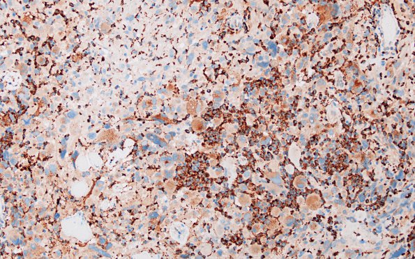
Washington University Experience | NEOPLASMS (GLIAL) | Pleomorphic Xanthoastrocytoma (PXA) | 3G PXA, anaplastic (Case 3) CD68 2
CD68 immunoreactivity labels macrophages, scattered microglia and, minimally, the cytoplasm of large pleomorphic cells. ---- Ancillary findings (not shown): No significant reactivity is observed for CAM 5.2, Melan-A, HMB-45, IDH1 (p. R132H) or BRAF V600E. Nuclear reactivity for ATRX is retained in the vast majority of the tumor cells. Reactivity for CD34 highlights blood vessels only. Periodic acid Schiff histochemical stain highlights eosinophilic granular cytoplasm. ---- Fluorescence in situ hybridization (FISH) studies found polysomies of chromosomes 7, 9, and 10, and no evidence of EGFR gene amplification, loss of 10 q/monosomy 10, or deletion of CDKN2A/monosomy 9. ---- Comment: The histomorphological, immunohistochemical and cytogenetic features of this high grade astrocytoma – particularly the marked pleomorphism, the presence of EGBs and vacuolated giant cells, the solid/circumscribed growth pattern, and the lack of monosomy 10/10q loss (reported in 75-95% of glioblastoma) – are most consistent with the diagnosis: Anaplastic pleomorphic xanthoastrocytoma, WHO Grade 3. Immunohistochemistry suggests that this example lacks the BRAF V600E mutation that has been reported in half or more of such cases. ---- Ancillary molecular testing includes: MGMT methylation not detected. Foundation One: PTEN exon 1&2 loss; TP53 1255T mutation ---- Diagnosis: Anaplastic Pleomorphic Xanthoastrocytoma, WHO Grade III

