Table of Contents
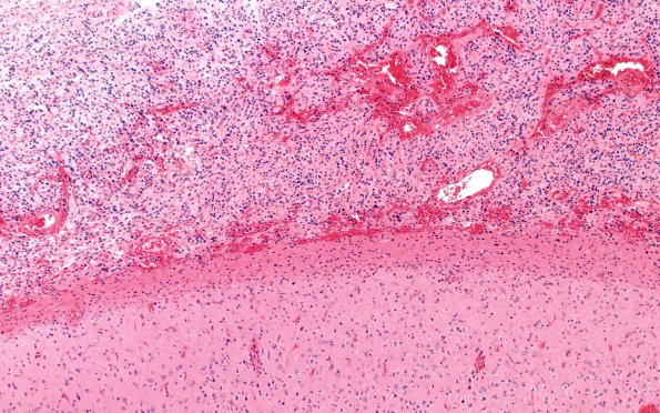
Washington University Experience | NEOPLASMS (GLIAL) | Pleomorphic Xanthoastrocytoma (PXA) | 4A1 PLeomorphic xanthoastrocytoma (Case 4) H&E B3 10X
Case 4 History
The patient is a 14 year old boy with new onset of seizures. Imaging shows a T2 abnormality in the left temporal lobe with enhancing contrast. Operative procedure: Craniotomy and excision of lesion.
4A1-5 This is a hypercellular neoplasm with a solid and infiltrative growth pattern and subarachnoid localization. The cells have round to elongated, large, pleomorphic, hyperchromatic nuclei with abundant foamy fibrillary, eosinophilic cytoplasm arranged as fascicles and infiltrating the adjacent cortex. Eosinophilic granular bodies are present and there is focal perivascular lymphocytic cuffing. Mitoses are hard to find.

