Table of Contents
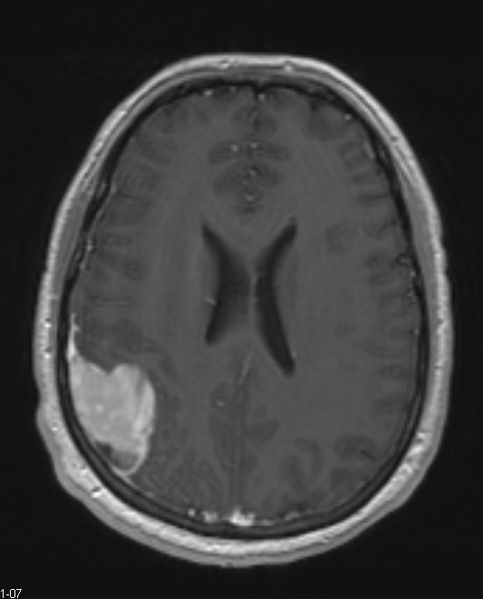
Washington University Experience | NEOPLASMS (GLIAL) | Pleomorphic Xanthoastrocytoma (PXA) | 9A Xanthoastrocytoma, pleomorphic (Case 9) T1 W 1 - Copy
Case 9 History ---- The patient is a 25 year old man who presented initially to an outside hospital after an episode of generalized tonic-clonic seizure. The patient underwent MR imaging, which showed an extra-axial mass in the right parietal region with a broadbased attachment to the dura. The mass was heterogenous with cystic areas, hypointense on T1, slightly hyperintense on T2, and enhanced avidly. There is a CSF cleft separating it from the underlying brain tissue. Operative procedure: Right parietal craniotomy and tumor resection. ---- 9A1,2 MRI studies show T1-weighted contrast administered enhanced axial and sagittal scans.

