Table of Contents
Washington University Experience | NEOPLASMS (GLIAL) | Pleomorphic Xanthoastrocytoma (PXA) | 9B1 Xanthoastrocytoma, pleomorphic (Case 9) H&E 1.jpg
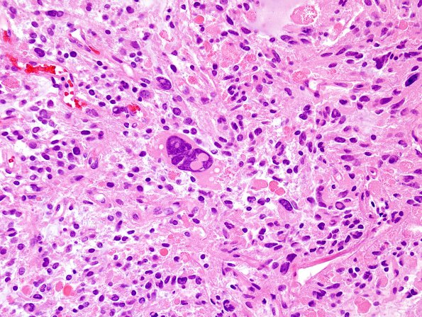
9B1-6 The tumor is arranged with a variable architecture. The cells display a wide range of morphologies, from those with small, bipolar, elongated nuclei and eosinophilic processes to large, multinucleated, highly pleomorphic cells with markedly atypical, giant nuclei. Frequent intranuclear inclusions are identified. There are numerous eosinophilic granular bodies (EGBs). There are cord-like, glandular and microcystic structures, as well as areas of more solid, sheet-like growth. Mitotic activity is rare. The tumor is richly vascularized, with a number of hyalinized vessels. The tumor appears to be closely apposed to the dura in areas. There is reactive meningothelial hyperplasia in arachnoid cap cells in multiple sections of dura adjacent to the tumor. The tumor does appear to infiltrate areas of cerebral cortex.

