Table of Contents
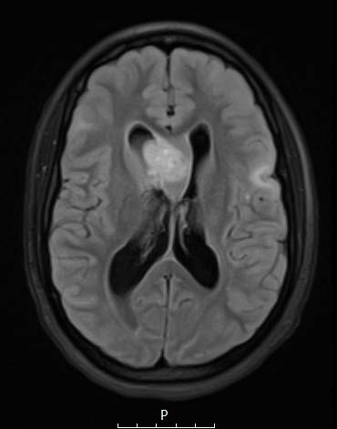
Washington University Experience | NEOPLASMS (GLIAL) | Subependymal Giant Cell Astrocytoma (SEGA) | 11A1 SEGA, in TS (Case 11) FLAIR 2 - Copy
Case 11 History ---- The patient was a 16-year-old girl with a history of tuberous sclerosis with epilepsy and mental retardation. She recently presented with a headache, photophobia and phonophobia. MRI showed multiple subependymal nodules and subcortical T2/FLAIR hyperintensities. There was a 3.6 cm mass at the foramen of Monro. Some areas of hemorrhage and calcification were noted in the mass. Radiologically, it was considered consistent with a subependymal giant cell astrocytoma. Operative procedure: Craniotomy for right frontal tumor. ---- 11A1-3 Imaging studies: 11A1 Prominent hyperintensity is seen in this FLAIR scan.

