Table of Contents
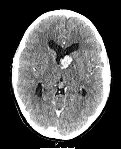
Washington University Experience | NEOPLASMS (GLIAL) | Subependymal Giant Cell Astrocytoma (SEGA) | 13A1 (Case 13) CT - Copy
Case 13 History ---- The patient was a 14-year-old girl with tuberous sclerosis complex and seizure disorder. MRI showed an enhancing mass in the left lateral ventricle near the foramen of Monro thought to represent a SEGA and cortical and subcortical tubers. The lateral ventricles were minimally enlarged. Operative procedure: Monteris ROSA laser ablation of left subependymal giant cell astrocytoma with external ventricular drain placement. ---- 13A1 A CT scan demonstrates a SEGA.

