Table of Contents
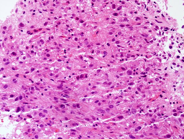
Washington University Experience | NEOPLASMS (GLIAL) | Subependymal Giant Cell Astrocytoma (SEGA) | 13B SEGA (Case 13) H&E 3.jpg
H&E stained sections of the biopsied enhancing intraventricular tumor show a low-grade astrocytic neoplasm. Neoplastic cells are arranged in moderately hypercellular sheets made up of short haphazardly oriented intersecting fascicles. There is no microvascular proliferation or necrosis. Neoplastic cells have spindled to gemistocytic morphology, moderate to focally marked nuclear atypia but no significant mitotic activity.

