Table of Contents
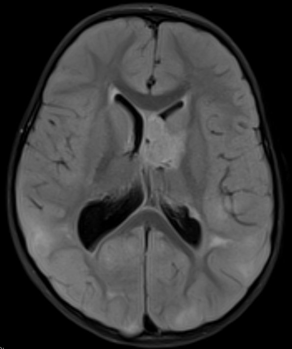
Washington University Experience | NEOPLASMS (GLIAL) | Subependymal Giant Cell Astrocytoma (SEGA) | 1A1 SEGA (Case 1) FLAIR 2 - Copy
Case 1 History ----- The patient was a 4-year-old girl with tuberous sclerosis, who presented with a brain mass near foramen of Monro. Operative procedure: IMRI craniotomy with tumor excision. ---- 1A1-4 MRI Examination
1A1 FLAIR scan shows the typical hyperintense appearance of a SEGA.

