Table of Contents
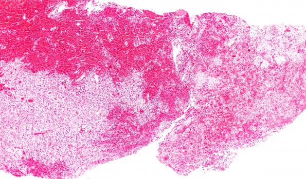
Washington University Experience | NEOPLASMS (GLIAL) | Subependymal Giant Cell Astrocytoma (SEGA) | 4A1 SEGA (Case 4) H&E 4X
Case 4 History ---- The patient was a 14 year old boy without any dermatologic stigmata of neurological disease, who presented following a seizure in April of 2013. MRI showed an enhancing T2-hyperintense 1.9 x 0.8 cm right frontal intraventricular mass. Radiological differential diagnosis included central neurocytoma, astrocytoma, oligodendroglioma, subependymal giant cell astrocytoma or, less likely, subependymoma. Operative procedure: Transcallosal resection of the intraventricular brain tumor with intraoperative MRI.
4A1-3 Hematoxylin and eosin stained sections of the neurosurgical specimen show many fragments of a low grade neoplasm with two distinct histological patterns. The dominant pattern (4A2) is characterized by large epithelioid cells with glassy eosinophilic cytoplasm, one or two eccentrically placed round-to-oval nuclei, and prominent nucleoli. In areas more classic for SEGA, these cells appear amongst smaller plump spindled cells with moderate eosinophilic cytoplasm and elongate oval nuclei. Microcalcifications are focally exuberant. There is no significant mitotic activity, pseudopalisading necrosis or microvascular proliferation.

