Table of Contents
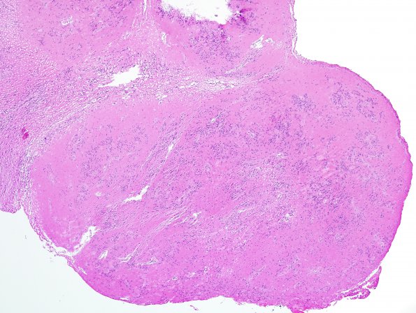
Washington University Experience | NEOPLASMS (GLIAL) | Subependymoma | 14A1 Subependymoma (Case 14) H&E 7
14A1-3 The provided H&E stained sections prepared from specimen #1 show several small fragments of classic subependymoma, characterized by poorly regimented lobules or clusters of cytologically bland cells with round-to-oval nuclei, relatively open chromatin and inconspicuous nucleoli, scattered within an expanse of dense fibrillar matrix. Mitotic figures, vascular proliferation and necrosis were not identified. ---- GFAP immunohistochemistry was strongly reactive in cell bodies and fibrillary matrix. Reactivity for epithelial membrane antigen (EMA) stained paranuclear dot-like structures. Ki-67 stained only very rare tumor cell nuclei (far fewer than 1%).This histological/immunohistochemical pattern of specimen #1 is diagnostic for subependymoma, WHO grade I

