Table of Contents
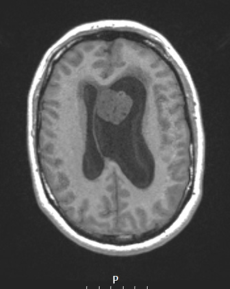
Washington University Experience | NEOPLASMS (GLIAL) | Subependymoma | 17A1 Subependymoma (Case 17) T1noC
Case 17 History ---- The patient is a 44yo male who presented with 8-12 weeks of right-sided weakness and ataxia, short term memory loss, and headaches. MRI showed a 2.9 cm intraventricular mass abutting the septum pellucidum with obstructive hydrocephalus of the lateral ventricles, thought clinically to represent central neurocytoma, with additional differential diagnoses including glial neoplasm and metastatic disease. Operative procedure: Bilateral craniotomy for resection of intraventricular mass ---- 17A1,2 The T1-weighted image without (17A1) and with (17A2) administered contrast shows a neoplasm abutting the septum pellucidum. Note the marked asymmetric hydrocephalus.

