Table of Contents
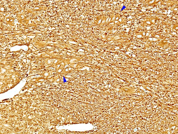
Washington University Experience | NEOPLASMS (GLIAL) | Subependymoma | 1C Subependymoma, spinal (Case 1) GFAP 4 copy
An immunohistochemical stain for GFAP shows strong positivity in neoplastic cells which are distributed in clumps. Ki-67 showed a labeling index of 4.1%, greater than typically seen in subependymomas. (GFAP IHC)

