Table of Contents
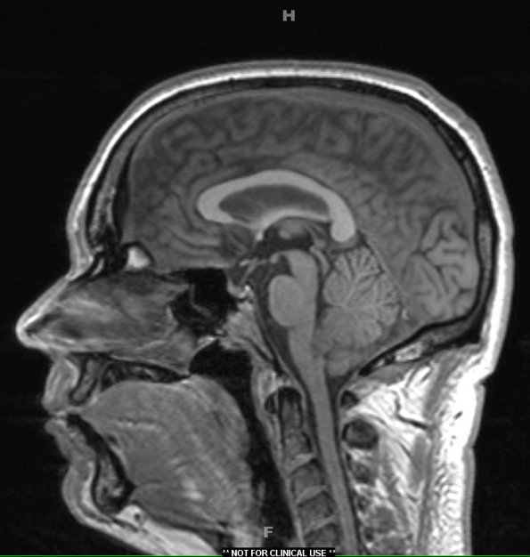
Washington University Experience | NEOPLASMS (GLIAL) | Subependymoma | 6A1 Subependymoma (Case 6) T1 1 - Copy
Case 6 History ---- The patient was a 40-year-old man found to have a brainstem tumor. An MRI showed a 1.2 cm T2-hyperintense non-enhancing lesion near the foramen of Magendie with mass effect on the posterior medulla. The radiographic differential included subependymoma, ependymoma and choroid plexus papilloma. Operative procedure: Posterior fossa craniotomy and tumor resection.---- 6A1,2 The subependymoma in this patient is small and extends to the obex as shown in this T1-weighted image (6A1) and as a iso/hyperintense neoplasm in this TIRM preparation (6A2).

