Table of Contents
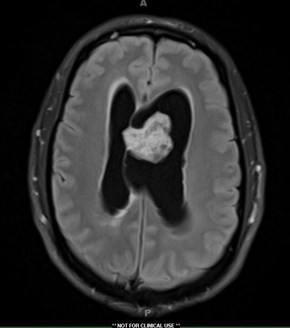
Washington University Experience | NEOPLASMS (GLIAL) | Subependymoma | 7A1 Subependymoma (Case 7) TIRM 1 - Copy
7A1-3 MRI showed a 3.2 x 3.1 x 4.2 cm mass with small internal cystic spaces within the left lateral ventricle with attachment to the septum pellucidum. The corpus callosum is thinned and both of the lateral ventricles are expanded. The mass is T-1 isointense, T2/FLAIR hyperintense, non-diffusion-restricted, heterogeneously enhances with contrast, and shows heterogeneous signal on susceptibility weighted imaging. ---- A TIRM sequence demonstrates pronounced hyperintensity.

