Table of Contents
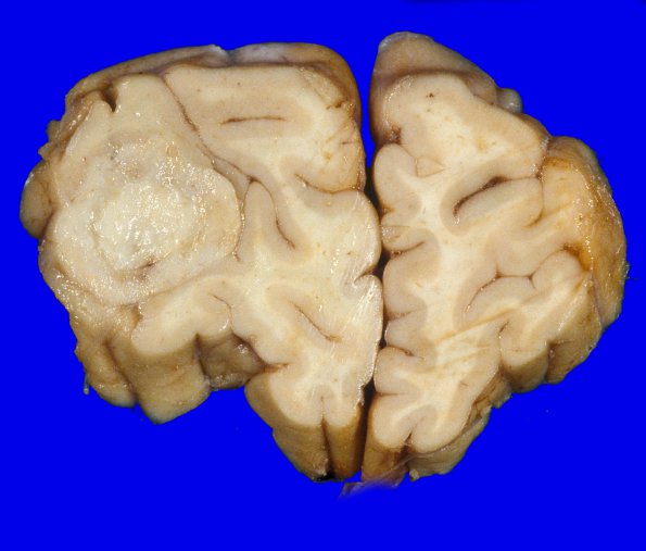
Washington University Experience | NEOPLASMS (METASTASES) | Gross Pathology | 18A1 Metastasis, lung (Case 18) 5
Case 18A1-6 Sections of the right frontal, left frontal, and left occipital lobe masses show poorly differentiated, metastatic adenocarcinoma most likely of lung origin with sharp brain-tumor interfaces, peripherally located microvascular proliferation and gemistocytic astrocytosis, macrophage infiltration, central, geographic necrosis, and single cell necrosis.

