Table of Contents
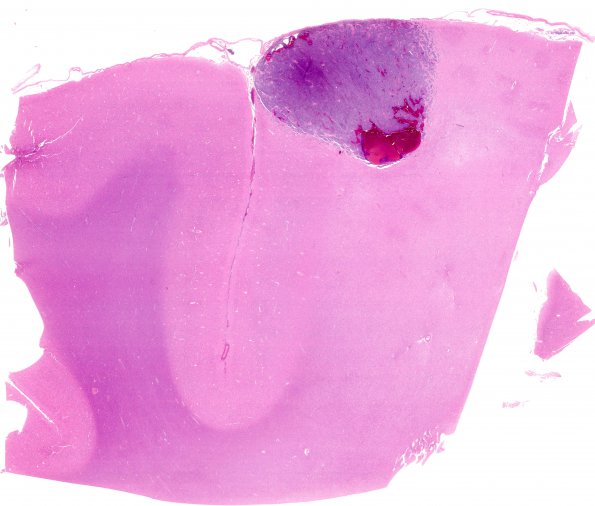
Washington University Experience | NEOPLASMS (METASTASES) | Melanoma | 2C1 Metastases, melanoma (Case 2) H&E whole mount
2C1-5 Sections show a well-circumscribed, neoplastic proliferation of predominantly spindled, but some epithelioid, cells arranged in vague fascicles and characterized by minimal-moderate amounts of eosinophilic cytoplasm, round to ovoid to markedly irregular and pleomorphic nuclei, scattered nuclear grooves, rare intranuclear inclusions, and coarse chromatin. Some small groups of cells as well as single cells can be seen invading the adjacent parenchyma. Mitotic figures are readily identified. There are scattered cells with intracellular, granular, brown pigment, consistent with melanin, as well as extracellular pigment. Areas of pooled blood are associated with the lesion, as well as hemosiderin pigment. The surrounding brain parenchyma shows focal necrosis (2C4) and reactive changes, including gliosis and mild edema. The histomorphologic appearance is consistent with that of a malignant melanoma. Sections of the left fronto-parietal cortical lesion show tumor at the gray-white junction with abundant associated hemorrhage

