Table of Contents
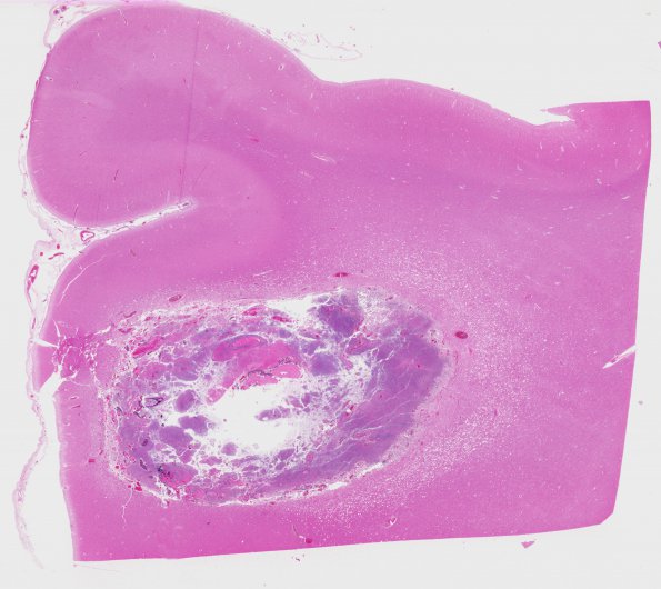
Washington University Experience | NEOPLASMS (METASTASES) | Meningeal | 13B1 Metastasis, lung (Case 13) H&E WM
13B1,2 Sections of the cerebral cortex show slight meningeal thickening. Sections from the lesions show metastatic carcinoma. Much of the tumor is necrotic. Where viable tumor cells are present they are arranged in small clusters and sheets, are epithelioid with scant cytoplasm, and show moulding in places and contain hyperchromatic nuclei. Mitoses are frequent. The morphological findings are consistent with a small-cell carcinoma. In addition, the adjacent brain parenchyma around these lesions shows exuberant gliosis and patchy vacuolation involving both the cortex and white matter.

