Table of Contents
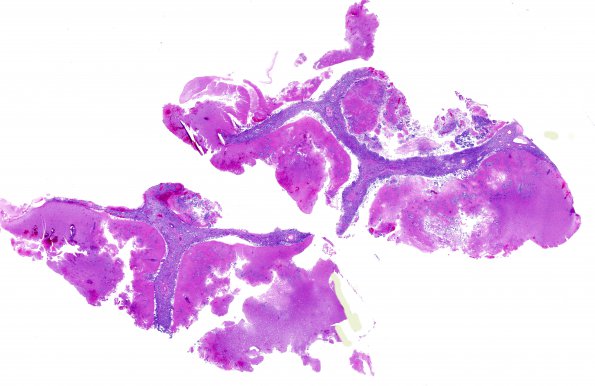
Washington University Experience | NEOPLASMS (METASTASES) | Meningeal | 16A1 Metastasis, lung, lepto (Case 16) H&E WM
16A1-3 Sections show partially necrotic brain tissue that is infiltrated by a sheet of tumor cells with a tubular and papillary growth pattern in a myxoid stromal background. There is marked leptomeningeal carcinomatosis with tumor cells, involving the pia and extending down into the brain parenchyma through the Virchow Robin spaces.

