Table of Contents
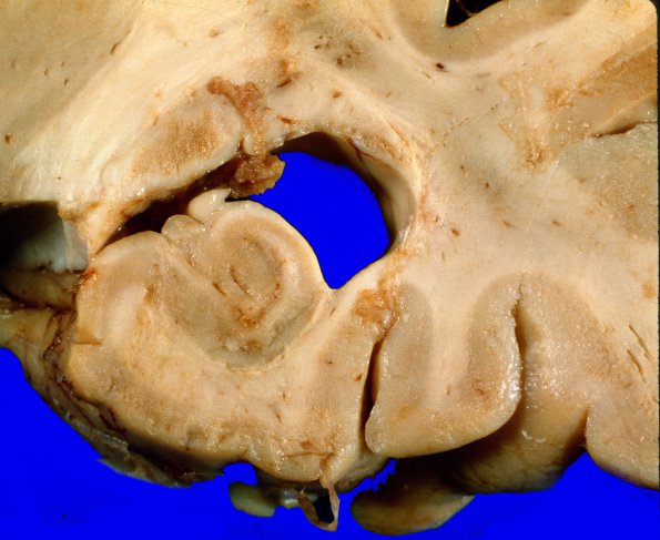
Washington University Experience | NEOPLASMS (METASTASES) | Meningeal | 1A8 Metastases, Lung primary (Case 1) X 10
The cerebral hemispheres are abnormal throughout their extent. This abnormality consists of multiple foci of ill-defined, granular white discoloration. These lesions vary up to approximately l cm. in greatest dimension, although most are rather ill-defined. Others possess a more distinct outline. These lesions are located both in grey and white matter, although they predominate in the cortex and at the grey-white junction. The grey matter of the cerebral hemispheres, the basal ganglia, brainstem and the cerebellum are extensively involved. Metastatic deposits were also present in the cerebral white matter and spinal cord.

