Table of Contents
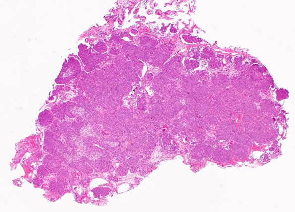
Washington University Experience | NEOPLASMS (METASTASES) | Microscopic | 15A1 Metastasis, medullary thyroid (Case 15) H&E 1 - Copy
Case 15 History ---- The patient is a 38-year-old man with a history of medullary thyroid carcinoma with lung and cerebellar metastasis. He underwent fractionated radiation to the sellar/suprasellar metastasis in June 2015. He developed blurry vision and underwent a transsphenoidal surgery for pituitary tumor resection in September 2018 at an outside hospital. His vision initially improved but later worsened. Brain MRI in 12/2018 showed a partially debulked sellar/suprasellar mass that has increased in size compared to the exam from 9/2018. Operative procedure: Endoscopic transsphenoidal hypophysotomy ---- 15A1,2 Routine H&E stain showed a malignant neoplasm with predominantly solid growth pattern arranged in large nests. Focal glandular formations are also appreciated. The tumor cells are predominantly spindled with pleomorphic nuclei and moderate amount of eosinophilic cytoplasm. Brisk mitotic activity and central comedo like necrosis are seen.

