Table of Contents
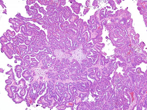
Washington University Experience | NEOPLASMS (METASTASES) | Microscopic | 16A1 Metastasis, papillary thyroid CA (Case 16) H&E - Copy (4)
Case 16 History ---- The patient is a 58-year-old man with a history of metastatic papillary thyroid carcinoma. He was initially diagnosed and treated at an outside hospital. He underwent thyroidectomy with lymph node dissection; those specimens reportedly showed papillary thyroid cancer with lymph node metastases. He was subsequently treated with radioactive iodine (2000). He later underwent left upper lobe and left lower lobe wedge resections showing metastatic papillary thyroid carcinoma. He recently presented with new onset headaches, nausea, and vomiting. Brain MRI in 3/2016 shows an enhancing, hemorrhagic lesion in the left cerebellar hemisphere and a second enhancing lesion in the lateral precentral gyrus with associated microhemorrhage. Radiological impression: Favor metastases. Operative procedure: Left posterior fossa craniectomy and resection of left cerebellar metastases with stereotactic navigation. ---- 16A1-3 H&E shows multiple fragments of a neoplasm forming true papillae with fibrovascular bundles. Generally appearing within a columnar epithelium, the tumor cells have eosinophilic cytoplasm, distinct cell borders, and basophilic nuclei with powdery chromatin. Occasional cells have cleared-out chromatin, nuclear grooves, and nuclear pseudoinclusions.

