Table of Contents
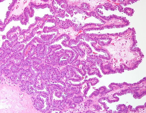
Washington University Experience | NEOPLASMS (METASTASES) | Microscopic | 16A3 Metastasis, papillary thyroid CA (Case 16) H&E 20X.jpg
H&E shows multiple fragments of a neoplasm forming true papillae with fibrovascular bundles. Generally appearing within a columnar epithelium, the tumor cells have eosinophilic cytoplasm, distinct cell borders, and basophilic nuclei with powdery chromatin.

