Table of Contents
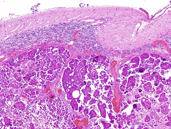
Washington University Experience | NEOPLASMS (METASTASES) | Microscopic | 18B1 Metastases, ovarian (Case 18) H&E 2.jpg
18B1-3 Sections of the right and left cerebellar tumor show nests of neoplastic cells forming micro-papillary structures. The neoplastic cells have abundant pink cytoplasm, with round hyperchromatic nuclei. Mitoses and necrosis are also seen. Numerous psammoma bodies are seen in the background.

