Table of Contents
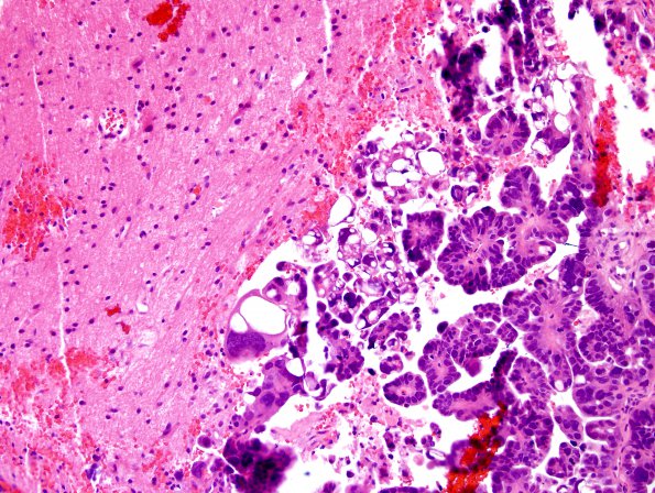
Washington University Experience | NEOPLASMS (METASTASES) | Microscopic | 19B Metastasis, ovarian mullerian CA (Case 19) H&E 1.jpg
There are multiple fragments of brain tissue containing islands of papillae and glands lined by large hyperchromatic neoplastic cells with a high nucleus-to-cytoplasm ratio and occasional intracytoplasmic mucin. Associated necrosis and calcifications are identified. This morphologic appearance is similar to the patient's previously diagnosed high grade serous adenocarcinoma involving the fallopian tubes and ovaries.

