Table of Contents
Washington University Experience | NEOPLASMS (METASTASES) | Microscopic | 1A2 Metastasis, colon primary (Case 1) H&E 2.jpg
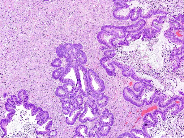
1A2,3 Higher magnifications of the tumor shown in image #1A1. The specimen consists predominantly of tumor arranged in glandular pattern with large areas of central necrosis. The tumor cells are mitotically very active, display moderate to severe pleomorphism with prominent nucleoli in most of the nuclei. (H&E)

