Table of Contents
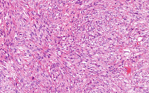
Washington University Experience | NEOPLASMS (METASTASES) | Microscopic | 26A1 Metastases, renal (Case 26) H&E 1
Case 26 ---- The patient is a 46-year-old man with a history of metastatic renal cell carcinoma of the right kidney with metastatic lesions to the brain (status post Cyber knife treatment in December 2017), lung, left hip, and right acetabulum. Operative procedure: Brain tumor resection. ---- 26A1,2 Sections show a metastatic poorly differentiated carcinoma with a relatively sharp demarcation between the tumor and the surrounding brain parenchyma which is surrounded by endothelial vascular proliferation. The tumor cells are epithelioid or spindled, with large nuclei, hyperchromatic chromatin, and prominent nucleoli. There are numerous mitotic figures, accumulation of foamy macrophages, hemosiderin/hematoidin deposition, and large areas of necrosis.

