Table of Contents
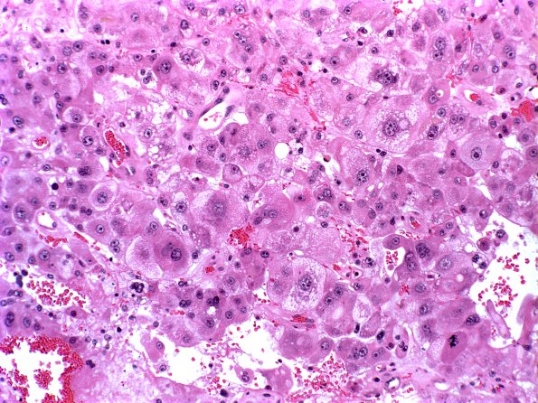
Washington University Experience | NEOPLASMS (METASTASES) | Microscopic | 2A Metastases Liver origin (Case 2)
Case 2 History ---- The patient is a 36 year old woman with a history of hepatocellular carcinoma, now with a brain tumor in the right frontotemporal area. ---- 2A Sections of the specimen show a metastatic neoplasm composed of cells with abundant eosinophilic to foamy cytoplasm and round to oval hyperchromatic nuclei with prominent nucleoli, arranged in cords, trabeculae or sheets. Mitotic figures and nuclear pleomorphism with multinucleation are frequently identified. ---- The neoplastic cells stain positive for alpha-fetoprotein and negative for cytokeratins 7 or 20. Both the histomorphologic features and immunohistochemical results support the diagnosis of metastatic hepatocellular carcinoma. CNS metastasis from hepatocellular carcinoma is rare (H&E)

