Table of Contents
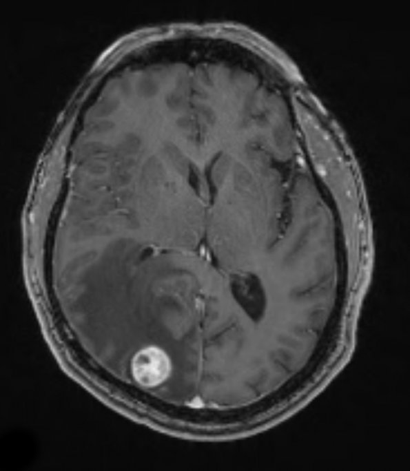
Washington University Experience | NEOPLASMS (METASTASES) | Microscopic | 34A Metastasis, renal (Case 34) T1 W 2
Case 34 History ---- The patient is a 59 year old man with a previous history of clear cell renal carcinoma. The patient presented with a history of headaches. ---- 34A MRI shows an enhancing T1-weighted contrast enhancing mass in the right occipital lobe. Operative procedure: Craniotomy with excision. ---- Not shown: The neoplasm had a pushing border and was well-circumscribed with a thin capsule. Numerous dilated vascular channels are present and intertwine between cords and nests of neoplastic cells. The neoplastic cells have well defined cytoplasmic membranes, show variably cleared cytoplasm, and contain irregularly-shaped round to ovoid nuclei with prominent nucleoli. Mitoses are present. The neoplastic cells are positive for CD10 and RCC. They are negative for CK7 and CD20.

