Table of Contents
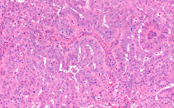
Washington University Experience | NEOPLASMS (METASTASES) | Microscopic | 51A1 Metastasis, giant cell lung CA (Case 51) H&E 6
Case 51 History The patient is an 82-year-old woman with severe headache, nausea/vomiting, and abdominal pain for three days. CT scan showed a 4.6 cm right paratracheal mass and bilateral adrenal nodules. Brain MRI showed a 4.1-cm right cerebellar intraparenchymal hematoma with a focus of enhancement. Operative procedure: Suboccipital craniotomy for resection of intra-axial right cerebellar hemorrhagic tumor. ----- 51A1-6 Routine H&E stained section of the "right cerebellar hemorrhage" show a poorly differentiated malignant neoplasm with abundant hemorrhage. In some areas the tumor has a epithelial and adhesive appearance. In others, the tumor cells are discohesive and have pleomorphic nuclei with abundant amount of pale eosinophilic cytoplasm. Frequent tumor giant cells are seen. There are abundant neutrophils within the background. Some tumor cells are seen to engulf red blood cells as well as neutrophils.

