Table of Contents
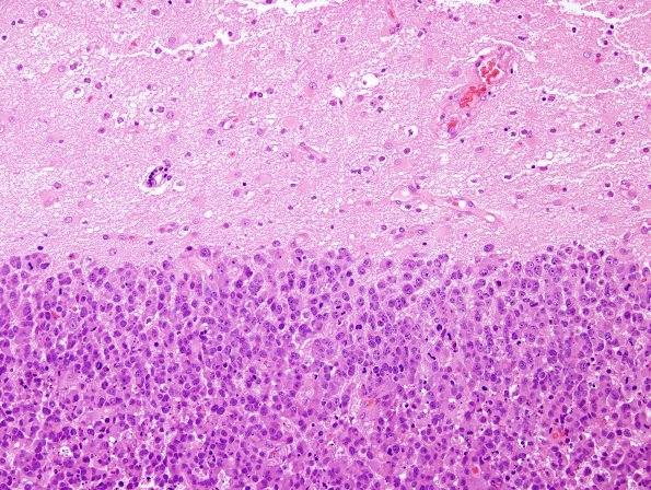
Washington University Experience | NEOPLASMS (METASTASES) | Microscopic | 52B1 Metastasis, large cell neuroendocrine CA (Case 52) H&E 11.jpg
52B1,2 Sections of the "right frontal tumor" show sheets and nests of neoplastic cells with abundant cytoplasm with round, hyperchromatic nuclei containing prominent nucleoli, and numerous mitoses. The neoplastic cells have sharp cell borders, with no intercellular bridges. Numerous apoptotic bodies are seen as well as necrosis.

