Table of Contents
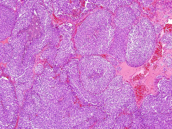
Washington University Experience | NEOPLASMS (METASTASES) | Microscopic | 56B1 Metastasis, lung, neuroendocrine carcinoma (Case 56) H&E 1.jpg
56B1,2 H&E stained sections of the intradural extramedullary tumor resection material show a peripheral nerve root expanded by a deposit of intra-neural metastatic carcinoma. The malignant cells are arranged in cohesive, highly cellular sheets that are partitioned by fibrovascular septae into variably sized lobules and large nests; some nests show central necrosis. The neoplastic cells are relatively uniform with oval nuclei, speckled chromatin suggestive of neuroendocrine differentiation, and modest amounts of cytoplasm. Mitotic activity is brisk.

