Table of Contents
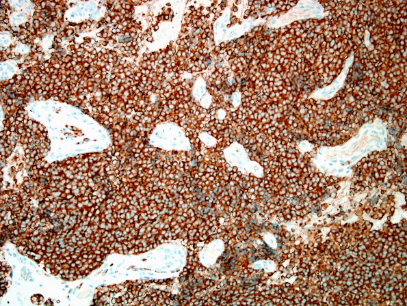
Washington University Experience | NEOPLASMS (METASTASES) | Microscopic | 56C Metastasis, lung, neuroendocrine carcinoma (Case 56) SYN 2.jp
There is strong reactivity within tumor cells for synaptophysin. ---- Not shown: The tumor stains strongly with CAM 5.2 and Chromogranin A and weak reactivity within a subset of tumor cell nuclei for TTF-1. Reactivity for S-100, collagen IV and neurofilament highlight the non-neoplastic components of the involved peripheral nerve; there is no evidence of sustentacular cells. Reactivity for proliferation marker Ki-67 (MIB-1 antibody) stains a large minority of tumor cells, estimated to be 25-35%. No significant tumor cell reactivity is observed for pancytokeratin (AE1/AE3), cytokeratin 7, cytokeratin 20, epithelial membrane antigen (EMA), progesterone receptor (PR), D2-40 (podoplanin), glial fibrillary acidic protein, S-100, collagen IV, or neurofilament. ---- Comment: These histologic and immunohistochemical features are most consistent with a high grade metastasis from the patient's previously diagnosed neuroendocrine tumor (atypical carcinoid) of the lung. The diagnoses of schwannoma, ependymoma, meningioma, and paraganglioma are effectively excluded. As part of this evaluation, the current slides were compared to those from the patient's primary lung tumor; the cytologic features are similar.

