Table of Contents
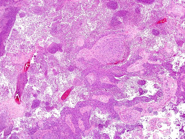
Washington University Experience | NEOPLASMS (METASTASES) | Microscopic | 64A1 Metastasis, lung & Marked Rxn (Case 64) D2 H&E 4
Case 64 History ---- The patient is a 63-year-old African American woman with a history of Grave's disease, hypertension, and psychosis who was recently admitted for a psychotic episode. Subsequent workup included head CT and MRI that showed a 1 cm avidly enhancing lesion in the left parieto-occipital region with adjacent vasogenic edema. Whole body PET shows increased FDG uptake in the left parieto-occipital mass, and right supraclavicular, right paratracheal, retrotracheal, right peribronchial, and right hilar lymphadenopathy. Excisional biopsy of a right supraclavicular lymph node showed metastatic carcinoma with a lung primary being favored (reactivity for AE1/AE3, CK7, TTF-1, and non-reactivity for CK20). Operative procedure: Craniotomy for tumor excision. ---- 64A1,2 Sections show a malignant neoplasm growing with a sheeted and nested architecture that contains numerous thickened irregularly shaped vessels. The neoplastic cells are predominantly epithelioid, have eosinophilic cytoplasm, and have round irregularly shaped nuclei with cleared chromatin and prominent nucleoli. Mitotic figures are easily identified. In multiple foci the neoplastic cells become highly discohesive. Marked inflammation is present and involves areas of nested neoplastic cells, much of its associated vasculature, and surrounding brain parenchyma.

