Table of Contents
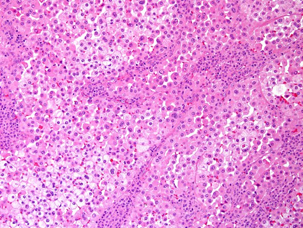
Washington University Experience | NEOPLASMS (METASTASES) | Microscopic | 65A1 Metastasis, lung, unusual (Case 65) H&E 4.jpg
Case 65 History ---- The patient is an 82-year-old woman with a history of lung adenocarcinoma, status-post left upper lobe lobectomy showing AJCC TNM stage T1a N0 MX disease (3/2014). She did not receive chemotherapy or radiation for her lung cancer. She has been followed with serial chest computed tomography (CT) scans, which have showed no evidence of progression or recurrence. She recently presented with difficulty speaking. MRI imaging on 03/2016 shows multiple enhancing, hemorrhagic lesions, the largest of which is in the left temporal parietal region. Radiological impression: Favor metastases. Operative procedure: Left craniotomy for tumor resection. ---- 65A1-H&E stained tissue shows a neoplasm with a broad brain/tumor interface. The tumor tissue is formed by lobules of relatively uniform cells, bordered by broad, cellular, irregular fibrovascular bundles. The tumor cells are epithelioid, with distinct cell borders, one-to-three large round nuclei with prominent nucleoli, and generous eosinophilic or partially cleared/vacuolated cytoplasm. On the intraoperative cytological smear preparation, the cells appear individually (i.e., are not cohesive). Mitotic figures are present but are not abundant.

