Table of Contents
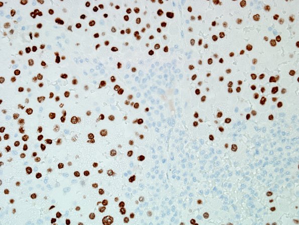
Washington University Experience | NEOPLASMS (METASTASES) | Microscopic | 65C Metastasis, lung, unusual (Case 65) TTF1 1.jpg
Immunohistochemically stained sections show strong reactivity for TTF1. ---- Not shown: The tumor cells are negative for Cytokeratin 20, Vimentin (fibrovascular bundles, only), Melan A, S100, Inhibin, and glial fibrillary acidic protein (GFAP). Neuron specific enolase (NSE) shows moderate staining in a subset of tumor cells. ---- In this clinical context, these histological and immunohistochemical findings support the diagnosis: Metastatic adenocarcinoma, consistent with lung primary.

