Table of Contents
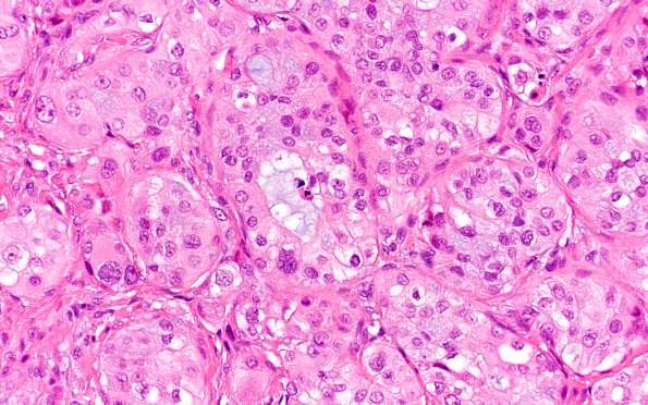
Washington University Experience | NEOPLASMS (METASTASES) | Microscopic | 66A1 Metastasis, lung mucoepidermoid (Case 66) 40X
66A1,2The tumor is arranged in large nests with solid, glandular, and cribriform architectures. The tumor cells are notable for both an "epidermoid" morphology, with relatively abundant amounts of eosinophilic cytoplasm, as well as mucinous differentiation in a majority of cells. Focally, there is clearing of cytoplasm within some of the more solid nests. The nuclei range in shape from round to markedly irregular, with patterns of chromatin distribution ranging from finely granular to vacuolated. Occasional prominent nucleoli are seen but are rare. Mitotic activity is noted. Occasional intranuclear inclusions are identified. In multiple areas cells infiltrate in smaller, more irregular clusters and singly. There is associated chronic inflammation. The tumor abuts bone, which appears reactive and remodeled. Individual tumor cell necrosis is identified, but the majority of the lesion is viable.

