Table of Contents
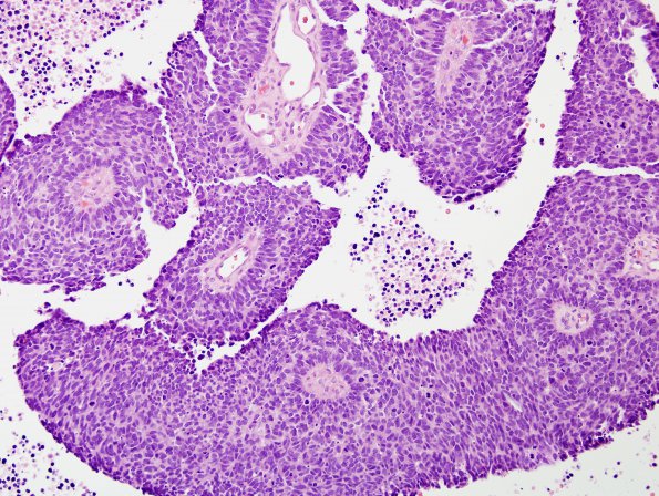
Washington University Experience | NEOPLASMS (METASTASES) | Microscopic | 67A1 Metastasis, lung small cell CA (Case 67) H&E 1 - Copy
67A1,2 Sections show a poorly differentiated, hypercellular proliferation of small, hyperchromatic cells with scant cytoplasm, nuclear molding, and numerous apoptotic bodies as well as geographic necrosis and frequent mitotic activity. The cells have very little visible cytoplasm and are growing in predominantly a sheet-like architecture, disrupted only by the numerous vessels throughout the tumor. Finally, there are scattered lymphocytes and plasma cells involving the adjacent dural/leptomeningeal tissue. Overall, the histomorphologic impression favors a small cell carcinoma.

