Table of Contents
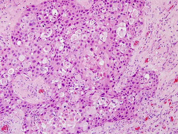
Washington University Experience | NEOPLASMS (METASTASES) | Microscopic | 69A Metastasis, lung squamous (p40 ) (Case 69) 2.jpg
Case 69 History ---- The patient is a 77 year old woman with a left occipital brain lesion. Operative procedure: Left occipital craniotomy for
resection of brain tumor with possible IMRI. ---- 69A Sections of the brain mass consists of nests of moderate to poorly differentiated squamous cell carcinoma with extensive necrosis which has the same histomorphology as the primary lung squamous cell carcinoma. Focal keratin formation is present.

