Table of Contents
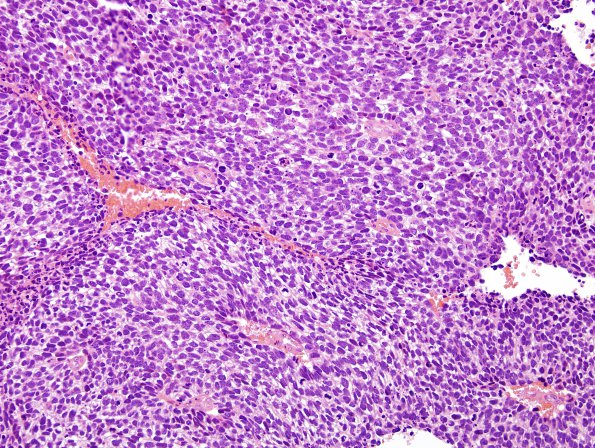
Washington University Experience | NEOPLASMS (METASTASES) | Microscopic | 71A1 Metastasis, lung, small cell CA (Case 71) H&E 1
Case 71 History ---- The patient is a 66 year old man with a left upper lobe lung lesion and liver metastasis that also has multiple solid and cystic enhancing masses in the cerebral hemispheres bilaterally with surrounding vasogenic edema. A large left frontal lobe cerebral mass measuring 6.6 cm is present causing subfalcine herniation. Operative procedure: Craniotomy for tumor resection. ---- 71A1,2 Sections show a hypercellular tumor centering around blood vessels with multiple areas of necrosis and blood between nests of tumor. The tumor cells have scant cytoplasm and show nuclei with a stippled ("salt and pepper") chromatin pattern and inconspicuous nucleoli. There is evidence of nuclear molding. There are numerous mitoses and frequent karyorrhectic debris.

