Table of Contents
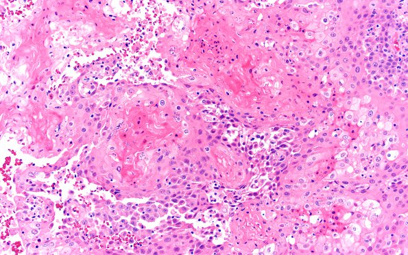
Washington University Experience | NEOPLASMS (METASTASES) | Microscopic | 73A1 Metastasis, lung, squamous (Case 73) H&E 20X 3
Case 73 History ---- The patient is a 68-year-old man with a history of stage III non-small cell lung cancer status post chemotherapy and radiation (MRI in 1/2021 negative for metastasis), who presented with confusion. MRI in April 2021 showed a 7 cm cystic lesion, now status post right frontal craniotomy for cyst evacuation and biopsy, however cultures were negative and OSH pathology did not reveal malignancy or other clear diagnosis. Repeat MRI (6/2021) showed a large, ring enhancing mass within the right frontal lobe, concerning for high-grade glioma. Operative procedure: Craniotomy excision tumor right frontal tumor resection. ---- 73A1,2 H&E sections show a metastatic carcinoma with keratinizing squamous differentiation. Some areas exhibit vague pseudopapillary features. The metastatic carcinoma demonstrates a pushing border with adjacent brain parenchyma. There is multifocal inflammatory infiltration at the carcinoma-brain interface.

