Table of Contents
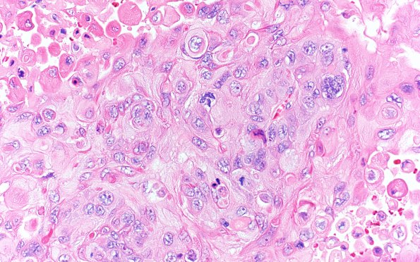
Washington University Experience | NEOPLASMS (METASTASES) | Microscopic | 74A Metastasis, lung, squamous (Case 74) H&E 40X
Case 74 History ---- The patient is a 63-year-old woman with a history of squamous cell lung carcinoma status post chemotherapy and resection (right upper lobectomy for recurrence in 11/2020), who presented with one week of short term memory and concentration difficulties. MRI showed a 4.6 cm peripherally enhancing left frontal lesion and 1.1 cm peripherally enhancing right frontal lesion concerning for metastatic disease with multifocal primary high grade glial neoplasm considered less likely. Operative procedure: Craniotomy excision intracranial mass. ---- 74A H&E shows a metastatic non-keratinizing squamous cell carcinoma. Marked pleomorphism, brisk mitoses and tumor necrosis are noted. Adjacent brain parenchyma demonstrates reactive astrocytosis.

