Table of Contents
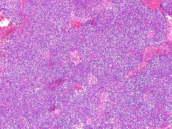
Washington University Experience | NEOPLASMS (METASTASES) | Microscopic | 75A1 Metastasis, lung, squamous (Case 75) H&E 3.jpg
Case 75 History ---- The patient is a 68 year old man with a history of metastatic lung cancer, who now presents with a 2.9 cm enhancing left frontal mass. Operative procedure: Left frontal craniotomy with tumor excision. 75A1,2 Sections of the left frontal lobe mass show sheets of cohesive tumor cells with multiple areas of keratinization and other areas that appear almost basaloid with nuclear palisading. The tumor cells have clear to eosinophilic cytoplasm and pleomorphic nuclei with clumped chromatin and variably prominent nucleoli. Scant glial tissue is seen. The morphologic findings are those of a metastatic squamous cell carcinoma and are consistent with a lung primary.

